
Radiation Biology (4/5): How ionising radiation is measured

Ionising radiation can cause sicknesses. In order to protect the population and personnel working in nuclear installations from these harmful effects, it must be possible to measure radiation doses at any time and as accurately as possible.
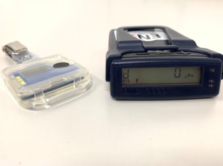
Safety is a top priority for any work conducted in nuclear installations. This is not only true for the operation of the installations, but also for protecting staff working there. Therefore, people who are considered to be exposed to radiation during their work in nuclear installations must wear, or carry, two different dose measuring instruments, so-called dosimeters. The dosimeters are regularly analysed and the accumulated doses are stored. The passive, approved dosimeter measures the radiation exposure for the central dose register. The active, electronic dosimeter also has an additional alarm function. It is only this function which makes it possible to intervene before personnel are exposed to excessive doses. If the statutory limit value for radiation exposure is exceeded, the regulatory authority decides in accordance with Art. 59 of the Radiological Protection Ordinance, whether the person concerned is to be placed under medical supervision.
If the dose is exceeded, the effective dose must be determined on an individual basis. In such a case, ENSI arranges for the dose to be recreated by external experts from the IRA (Institut de radiophysique) in Lausanne. In addition, a laboratory chromosome analysis is arranged (see section on biological dosimetry) as an additional safeguard. The biological dosimetry data are communicated to ENSI and the Swiss Accident Insurance Fund (Suva).
On this basis, the regulatory authorities take the necessary measures, such as the temporary or permanent exclusion from work as a classified radiation worker. Since 2009, there have been three cases where thresholds have been exceeded within ENSI’s oversight area. Thus far, no health effects have been observed for those affected.
Exceeding threshold values concerning nuclear power plant (NPP) employees in Switzerland
In recent years there have been three cases of a threshold being exceeded in Swiss nuclear installations:
- During the overhaul of the Beznau 2 NPP in August 2009, two employees were exposed to a dose of 37.8 mSv and 25.4 mSv respectively, while working below the reactor pressure vessel. This was caused by inadequate overhaul planning and deficiencies in the area of occupational radiation protection. In its assessment of the incident, ENSI concluded that, given the doses concerned, no direct health consequences were to be expected.
- In August 2010, a diver picked up an unknown object in the fuel element transfer basin during maintenance work at the Leibstadt NPP. In doing so, the radiation exposure to his hand exceeded the permissible limit. The estimation of the radiation exposure resulted in a dose value of up to 7.5 Sv (skin dose) – for classified radiation workers the skin dose limit is 500 mSv per year. To date, no resulting health consequences have been identified.
What is biological dosimetry?
Biological dosimetry is used when no physical measuring method is available or when the results of such measurements are in doubt. It can also be considered as a supplement to the dosimeter and it is particularly important if suspected radiation exposure has to be confirmed retrospectively. Thanks to biological dosimetry, it is possible to investigate how a given (high) radiation dose affects a cell. Not only can radiation-induced changes be detected, but it is also possible to draw conclusions about the extent of the dose.
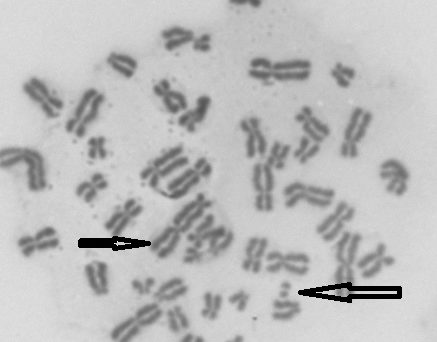
In biological dosimetry, metabolic elements or biological structures in the human body, or their changes after irradiation, are used as a “dosimeter”. The analysis of dicentric chromosomes is regarded as the global gold standard, especially in cases concerning the detection of acute whole-body radiation exposure. These are chromosomes that have had their form changed by irradiation and which are visually easy to analyse because they have two easily-recognisable constrictions (centromeres). Undamaged chromosomes only have one centromere, which divides the chromosome into two arms.
In a chromosome analysis, cells from blood samples taken from the arm vein are examined. The analysis can be performed manually using light microscopy or can also be automated. Technically, the evaluation is not very demanding, but it is time-consuming: for statistical reasons very many, at least 1,000, prepared cells are required so that it is also possible to detect lower doses. With the corresponding number of evaluated cells, doses up to significantly less than 100 mSv can be detected. The type of radiation can also be determined using calibration curves. In addition, these chromosomal changes are radiation-specific, i.e. they do not occur in the normal metabolism or, for example, in response to chemical contact. This allows us to answer the crucial question: “Did a radiation event occur or not?”
This is the fourth of five articles on radiation biology. The last article deals with the latest aspects of radiation biology.